Inverted microscope
MBL3200 - Transmitted light microscope for biology and medicine

Inverted microscope MBL3200
Laboratory microscope with extensive equipment
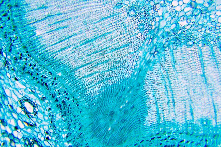
The MBL3200 inverse microscope is specially designed for the identification and analysis of biological substances and cultures. The objectives have a large working distance, making it possible, for example, to examine samples through the bottom of petri dishes. With an upright microscope, the lens is under the specimen and the condenser above the specimen, which is particularly suitable for viewing on microscope slides. In an inverted microscope, this arrangement is reversed. This gives the user more space and ensures the required proximity of the lens to the specimen, so that even living cells in cell culture dishes or other, larger containers can be easily analyzed. In addition, an inverted microscope allows easy access to the cells, for example micromanipulators. The photo and C-mount video adapter allows connection of camera and video camera.
- Large working distance
- Observation in larger containers, e.g. cell culture dishes possible
- Living cells in cell culture dishes or other, larger containers can be analyzed
- Documentation via connection for camera and video camera possible
- Wide range of accessories available
Typical applications
- For biology and medicine, testing laboratories and environmental institutes
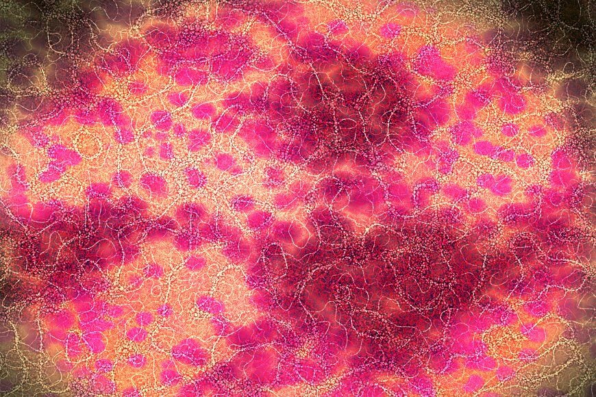
- For the identification and analysis of biological substances and cultures
- Identification of different cell structures
- For the investigation of living cells
- For examination in cell culture dishes and other large containers
- Possibility of micromanipulation
Model specifications
| MBL3200 | |
|---|---|
| BASIC EQUIPMENT | |
| OPTICAl HEAD | Inclined optical head,
Symmetrical eye distance adjustment (55 – 75 mm), Dioptre compensation with scale. |
| OBJECTIVE REVOLVER | Quadruple |
| OBJECTIVES | Planachromatic, infinity
4x/NA 0.10 // object field Ø: 5.5 mm 10x/NA 0.25 // object field Ø: 2.2 mm 40x/NA 0.65 // object field Ø: 0.55 mm PH20x/NA 0.40 // object field Ø: 1.1 mm |
| MAXIMUM OBJECT HIGHT | 24 mm with 4x/NA 0.10
23 mm with 10x/NA 0.25 21 mm with 40x/NA 0.65 |
| EYEPIECES | 10x plane eyepiece
Visual field number: 22 |
| CONDENSER | Brightfield condenser |
| ILLUMINATION | 6 V 30 W, adjustable |
| XY STAGE | Movement Range |
| STAND | Made of metal with coaxial coarse/fine knob |
| FURTHER EQUIPMENT | Iris diaphragm
Filter holder Blue filter Green filter |
| POWER SUPPLY | 90–240 V |